Magnetic Resonance Imaging (MRI)
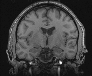
An MRI is a test that provides pictures of the inside of the body. A strong magnetic field is used to form these pictures, which is less harmful than X-rays and CAT scans. In patients with seizures, MRI images of the brain can sometimes show the cause of the seizures, such as a stroke, infection, tumor or abnormal brain tissue. By locating abnormal or damaged brain tissue, the MRI images can help pinpoint where in the brain seizures start, and surgeons may later be able to remove the damage and stop the seizures.
Because MRI uses a strong magnet, there are some special concerns. Some metals may be pulled into the magnet, so if you have metal in your body you may not be able to have an MRI. Be sure to tell your doctor if you have had spine or joint surgery, if you have a pacemaker, or if you know you have metal in your body. Also tell your doctor if you have had a job working with metal, such as welding.
The MRI procedure lasts about 45 minutes. You may get an injection of "contrast" dye that helps to spot some abnormalities. You will lie down on a table that carries you into the MRI machine, so tell your doctor before the MRI if you are claustrophobic. The MRI actually takes several different pictures of the brain, each of which shows the brain in a different way. Each complete picture takes about 5 minutes, and the technician will tell you when each picture starts and ends. It is very important to lie as still as possible while the MRI machine is on because even very small head movements can make the images blurry.