The Basics and What to Expect
X-Ray
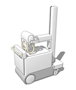
- Special type imaging that uses radiation called electromagnetic waves
- Creates pictures of inside your body when different tissues absorb different amounts of ionizing radiation
- These pictures demonstrate various shades of gray with air being black and bones being white
- X-rays can be harmful in large doses so we limit your exposure as best we can
- during the study a plate will be positioned underneath the area we are imaging and a tube opposite the plate will be used to take a picture
CT
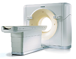
- Special type of imaging that uses multiple x-rays and computers to make very clear pictures of your body
- This technique again uses radiation; each slice of the scan shows a different level of your body
- During the study you lie still on a table that passes through the center of the larger machine
- Sometimes contrast is used like a dye in your veins to help improve images that we obtain
MRI
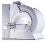
- Special type of imaging that uses magnets and radio waves to create pictures of the structures inside your body
- These pictures are created without the use of ionizing radiation
- Certain devices are not safe inside the large magnet and the technologist will check with you about them
- During the study you lie still on a table that slides inside a tunnel-shaped machine
- The machine may make a lot of noise while working
- Sometimes contrast is used like a dye in your veins to help improve images that we obtain
Ultrasound
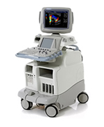
- Special type of imaging that uses high-frequency sound waves to create pictures of the structures in your body
- These pictures are created without the use of ionizing radiation
- During the study you lie on table while we use a handheld device called a transducer attached to a monitor/machine
- Pressing on the area of interest allows us to send sound waves into the body and capture the waves that bounce back
- We can also see flow of blood in vessels using sound waves that show up in color on the screen
Facts about Contrast
- We use contrast which is similar to a dye in order to obtain better images and provide your primary doctor with more useful information.
- Contrast is usually given either orally (by mouth) or intravenously (through an IV in your arm).
- Your body gets rid of the intravenous contrast through your kidneys, the more you urinate the quicker your body will remove the contrast from your blood.
- The risks of receiving contrast are allergic reaction (varying for very mild to severe) as well as worsening of kidney problems for those with pre-existing disease.
Imaging For Kids
- It is our goal to limit radiation exposure for our pediatric patients.
- MRI and US are frequently used as alternatives to standard imaging.
Imaging When Pregnant
- It is our goal to limit radiation exposure for our pregnant patients.
- MRI and US are frequently used as alternatives to standard imaging.
How to Prepare for your Examination
- Remember to bring a list of your medications
- Arrive early and check in as soon as possible
- Refrain from eating the morning of your examination
- Drink plenty of fluids after receiving a contrast enhanced examination
- Call well in advance for any changes or cancellations