
James B. Ranck, Jr, MD
Distinguished Teaching Professor Emeritus
Physiology and Pharmacology
Neural activity causes electrical currents that flow through the impedance of brain generating potential fields that we can record in our attempt to understand that activity. Also, variations in impedance shape the result when we pass our own currents to modify neural activity. Variations in impedance of brain areas, whether due to structure or behavior, affect current flows. My early interests were in how impedance related to these processes. I found that impedance of subiculum, part of hippocampal formation, changes substantially during sleep. This led me to wonder why it showed changes that varied with behavior. I found that the hippocampal formation has many fascinating aspects.
Since my discovery of the subicular impedance change during sleep, the research in my laboratory has focused on recordings from freely moving rats in brain areas that are involved in cognition, learning and memory. The brain areas of the hippocampal formation include hippocampus proper, subiculum, presubiculum, postsubiculum and entorhinal cortex. In the rat, much of the research is related to navigation, an easily quantified aspect of cognition, learning and memory. The principle cells of the hippocampus fire when the rat is in a circumscribed location in its environment; they have firing fields in a particular place on the floor, so are called “place cells”. Many neurons in entorhinal cortex have small firing fields that are arranged in a regular triangular grid pattern, so are called “grid cells”. They may be involved in measuring distance. My discovery of “head-direction cells” in the dorsal postsubiculum completed the set of three essential components of the rat navigational system: location, distance and direction. Each of these neurons fires rapidly when the rat’s head is pointed in a particular direction and is silent for other directions. All head directions are represented in the firing of different neurons among this population.
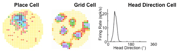
Figure 1. Examples of a hippocampal place cell, an entorhinal grid cell and a postsubicular head direction cell recorded as the rat foraged in a cylindrical chamber. For the place and the grid cell, higher firing rates are darker colors, yellow is zero.
In recent years I have been exploring an entirely new way to conceptualize the human intellect using rigorous topological principles. These topological rules can provide for analysis of the activity of populations of neurons, description of their products and fusion of the various aspects of that brain activity to ultimately describe the generation of consciousness. We have shown that the fundamental method works. The book is forthcoming.
- Ranck, J. B. (1973). Studies on single neurons in dorsal hippocampal formation and septum in unrestrained rats. 1. Behavioral correlates and firing repertoires. Exp. Neurol. 41, 462-531.
- Feder, R. and Ranck, J. B. (1973). Studies on single neurons in dorsal hippocampal formation and septum in unrestrained rats. 2. Hippocampal slow waves and theta cell firing during bar pressing and other behaviors. Exp. Neurol. 41, 532-555.
- Fox, S. E., and Ranck, J. B. (1975). Localization and anatomical identification of theta and complex spike cells in dorsal hippocampal formation of rats. Exp. Neurol. 49, 299–313.
- Fox, S. E., & Ranck, J. B. (1981). Electrophysiological characteristics of hippocampal complex-spike cells and theta cells. Exp. Brain Res. 41(3-4), 399–410.
- Muller, R. U., Kubie, J. L., and Ranck, J. B. (1987). Spatial firing patterns of hippocampal complex-spike cells in a fixed environment. J. Neurosci. 7, 1935–1950.
- Taube, J. S., Muller, R. U., and Ranck, J. B. (1990). Head-direction cells recorded from the postsubiculum in freely moving rats. I. Description and quantitative analysis. J. Neurosci. 10, 420–435.
- Taube, J. S., Muller, R. U., and Ranck, J. B. (1990). Head-direction cells recorded from the postsubiculum in freely moving rats. II. Effects of environmental manipulations. J. Neurosci. 10, 436–447.
- Quirk, G. J., Muller, R. U., Kubie, J. L., and Ranck, J. B. (1992). The positional firing properties of medial entorhinal neurons: description and comparison with hippocampal place cells. J. Neurosci. 12, 1945–1963.
- Muller, R. U., Ranck, J. B., and Taube, J. S. (1996). Head direction cells: properties and functional significance. Curr. Opin. Neurobiol. 6, 196–206.