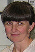
Sabina Hrabetova, MD, PhD
Professor
Department of Cell Biology
Sabina Hrabetova received an M.D. from Charles University in Prague in 1989, and an Ph.D. from the SUNY Downstate in 1998, working with Professor Todd Sacktor. She completed her postdoctoral training in Neuroscience and Biophysics at NYU School of Medicine under the mentorship of Professor Charles Nicholson. In 2002, Dr. Hrabetova began her independent research career as a Research Assistant Professor in the Department of Neuroscience and Physiology, NYU School of Medicine, directed by Professor Rodolfo Llinás. She joined the faculty of the Department of Cell Biology, SUNY Downstate as an Assistant Professor in 2006.
Research
The long-term goal of Dr. Hrabetova’s research is to construct and characterize a realistic three-dimensional model of the brain extracellular space (ECS), in order to predict the impact of structural changes on the transport of signaling molecules, nutrients and therapeutic agents. ECS comprises the narrow channels that separate brain cells. It is essential for normal brain function and influences many critical processes including intercellular signaling, nutrient delivery and neurotrophic effects. Significantly, the ECS also forms the final route for all drug delivery to brain cells. Once introduced into the ECS, substances are transported predominantly by diffusion because the ECS lacks any active transport mechanism. Physiological effects of the substances diffusing in the ECS are governed by their concentrations and their arrival times, which in turn depend on the ECS structure affecting the diffusion. To develop quantitative understanding of any of these diffusion-mediated processes, essential structural parameters of the complex ECS environment must be identified and characterized.
Service
Service on committees of SUNY Downstate Health Sciences University, College of Medicine, School of Graduate Studies, Department of Cell Biology
Reviewer for grant agencies and scientific journals
Biophysical Studies of Brain Extracellular Space
Dr. Hrabetova’s laboratory combines experiments measuring diffusion in brain tissue with mathematical modeling to characterize the structure of the ECS and how it is regulated. ECS forms approximately 20% of the brain volume, and it is filled with ionic solution and macromolecules of the extracellular matrix. The basic ECS structure is determined by a) the geometrical shape of neurons and glia, b) the width of the pores between the cells, and c) the extracellular matrix. Dr. Hrabetova’s team studies the role of these factors, and how they change during brain activity and in pathological states. The results of these studies are important both for understanding how altered ECS structure in neuropathological states disrupts the chemical traffic of the brain, and for designing effective strategies to deliver drugs in patients suffering from neurological disorders and brain tumors.
The extracellular space compartment is classically viewed as static and functionally less significant than neuronal and glial compartments. Recent studies from Dr. Hrabetova’s laboratory indicate the contrary, that the volume of the extracellular space compartment changes dynamically in response to neuronal activity and that these changes have important functional consequences. It is expected that her research, focused on extracellular space volume dynamism, the underlying molecular mechanisms, and the functional impact of volume changes, will transform the existing view of the extracellular space compartment as static and noncontributory, thus overcoming the existing barrier in our understanding of the extracellular space and its role in the CNS function and dysfunction.
- Fringuello AR, Colbourn R, Goodman J, Michelson HB, Ling DSF, Hrabetova S (2024). Rapid volume pulsations of the extracellular space accompany epileptiform activity in trauma-injured neocortex and depend on the sodium-bicarbonate cotransporter NBCe1. Epilepsy Res 201:107337. PubMed PMID: 38461594
- Naik A, Jensen V, Bakketun CB, Enger R*, Hrabetova S*, Hrabe J* (2023). BubbleDrive,
a low-volume incubation chamber for acute brain slices. Sci Rep 13:20005. PubMed PMID:
37973847
*Corresponding authors - Thevalingam D, Naik AA, Hrabe J, McCloskey DP, Hrabetova S (2021). Brain extracellular space of naked mole-rats expands and maintains normal diffusion under ischemic conditions. Brain Research 1771:147646. PubMed PMID: 34499876
- Colbourn R, Hrabe J, Nicholson C, Perkins M, Goodman JH, Hrabetova S (2021). Rapid volume pulsations of the extracellular space coincide with epileptiform activity in mice and depend on the NBCe1 transporter. Journal of Physiology 599:3195-3220. PubMed PMID: 33942325
- Sherpa AD, Guilfoyle DN, Naik AA, Isakovic J, Fumitoshi Irie, Yamaguchi Y, Hrabe J, Aoki C, Hrabetova S (2020). Integrity of white matter is compromised in mice with hyaluronan deficiency. Neurochemical Research 45:53-67. PubMed PMID: 31175541
- Hrabe J, Hrabetova S (2019)*. Time-resolved integrative optical imaging of diffusion
during spreading depression. Biophysical Journal 117:1783-1794. PubMed PMID: 31542225
*Featured as New and Notable: Pajevic S (2019). Exploring the dynamics of brain extracellular space. Biophysical Journal 117:1781-1782. - Hrabetova S, Cognet L, Rusakov DA, Nägerl UV (2018). Unveiling the extracellular space of the brain: From super-resolved microstructure to in vivo function. Journal of Neuroscience 38:9355-9363. PubMed PMID: 30381427
- Nicholson C, Hrabetova S (2017). Brain Extracellular Space: The Final Frontier of Neuroscience. Biophysical Journal 113:1-10. PubMed PMID: 28755756
- Sherpa AD, Xiao F, Joseph N, Aoki C, Hrabetova S (2016)*. Activation of β-adrenergic
receptors in rat visual cortex expands astrocytic processes and reduces extracellular
space volume. Synapse 70:307-316. PubMed PMID: 27085090.
*Featured as Cover story and Editor’s Choice. - Xiao F, Hrabe J, Hrabetova S (2015)*. Anomalous extracellular diffusion in rat cerebellum.
Biophysical Journal 108:2384-2395. PubMed PMID: 25954895
*Featured as New and Notable: Nicholson C (2015). Anomalous diffusion inspires anatomical insights. Biophysical Journal, 108:2091-2093.
Teaching and mentoring are key components of Dr. Hrabetova’s professional life. As a student, she was inspired by her teachers and mentors to pursue research and education. She is now striving to return back what she has received. Whether in the classroom or in the laboratory, her goal is to build on students’ motivation and create an environment that would help them to learn new material or to master a new experimental technique. Furthermore, her goal is to instill the values of science-driven medicine and hypothesis-driven research in the hearts and minds our students.
Dr. Hrabetova is teaching classes at the College of Medicine, School of Health Professions and School of Graduate Studies.