
Frank Scalia, PhD
Professor
Department of Cell Biology
Dr. Scalia, a graduate of New York University, received his PhD in Neuroanatomy at the Downstate Medical Center in 1964, where he built his reputation for studies on the structure and connections of the olfactory system, leading to his discoveries on the parallel but separate projections of the main olfactory and vomeronasal systems. Dr. Scalia contributed to the development of methodologies for the detection of the anterograde transport of HRP, both at the LM and EM level, and used this method for studies on the visual pathway in species as diverse as the rat and the frog. Combining degeneration and anterograde transport techniques in a positive-negative paradigm allowed fine analysis of topographic connections in the pretectal region for the first time, and clarified the previously ambiguous cellular organization of this part of the visual system. His work on the frog led to his interest in optic nerve regeneration. Notable among his early studies of optic nerve regeneration were the in vivo delineation of growth cone morphology, the observation that retinal ganglion cells move about after section of the optic nerve, and the discovery that regenerating retinal axons from the part of the retina that images the frontal visual field can form functioning synaptic connections in a specific region of the olfactory cortex. The last finding suggests that the emergence of this highly unusual projection is a neuroplastic phenomenon reflecting a morphological stabilization of otherwise labile synapses by means of a form of Hebbian reinforcement accompanying feeding. More recently, Dr. Scalia has turned his attention to the role of the Eph/ephrin families in the development and remapping of the retinotectal system. The imaging surface of the retina is mapped point for point onto the visual centers of the brain, providing the brain with a coherent view of the visual world. The formation of these visuotopic maps, which are essential for normal vision and its recovery after optic nerve regeneration, involves the independent mapping of the naso-temporal and dorso-ventral axes of the retina onto the antero-posterior and medio-lateral axes of the visual centers of the brain. His work in this area provided evidence that the EphA/ephrin-A family is involved in the mapping of the naso-temporal retinal axis in all vertebrates studied, but that the role of the EphB/ephrin-B family, which is partly involved in mapping the dorso-ventral axis in some species, is not conserved in all. In current work, Dr. Scalia has published a brain atlas of a tropical species of bat, which presents the first detailed and modern analysis of the thalamus and amygdala of these neglected mammals, and is studying the structure of its visual system in comparison with that of the domestic mouse.
Dr. Scalia is a member of the teaching staff responsible for the Neuroanatomy/Neuroscience curriculum for the Medical School and the School of Health Professions. His long-term and broad experience as a professional neuroanatomist has provided a valuable resource to junior staff over the years. He has authored many of the laboratory manuals, the first of which was copyrighted in 1972, and the current version of it remains in use in the Neuroscience block.
- Bach, H., Feldheim, D.A., Flanagan, J.G. and Scalia, F. Persistence of graded EphA/ephrin-A expression in the adult frog visual system. J. Comp. Neurol. 467:549-565, 2003.
- Bach, H., Arango, V., Feldheim, D.A., Flanagan, J.G. and Scalia, F. Fiber order of the normal and regenerated optic tract in the frog (Rana pipiens). J.Comp. Neurol. 477:43-54, 2004.
- Scalia, F. and Feldheim, D.A. Eph/ephrin A- and B- family expression patterns in the leopard frog (Rana utricularia). Dev. Brain Res. 158:102-106, 2005.
- Yagita, Y., Barjis, I., Hecht, M., Bach, H., Feldheim, D.A. and Scalia, F. Partial nucleotide sequences and expression patterns of frog (Rana pipiens)ephrin-A2 and ephrin-A5. Dev. Brain Res. 159:72-77, 2005 (cover article).
- Scalia, F., Currie, J.R. and Feldheim, D.A. Eph/ephrin Gradients in the Retinotectal System of Rana pipiens: Developmental and Adult Expression Patterns. J. Comp. Neurol. 513: 30-48, 2009.
- Higenell, V., Han, S.M., Feldheim, D.A., Scalia, F. and Ruthazer, E.S. Expression patterns of Ephs and ephrins throughout retinotectal development in Xenopus laevis. Dev. Neurobiol., 72:547-563, 2012.
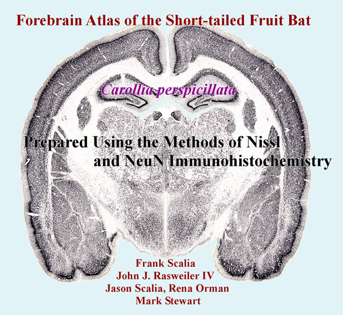
- Scalia, F., Rasweiler, J.J. IV, Scalia, J., Orman, R. and Stewart. M. Forebrain atlas of the short-tailed fruit bat, Carollia perspicillata. Prepared using the methods of Nissl and NeuN immunohistochemistry. Springer, 2013